.png)
Pituitary Lesion Vision Loss: When Sight Problems Start in the Brain
- Keshav Narain, M.D.
- Aug 11, 2025
- 3 min read
Updated: Aug 20, 2025
Not all vision problems start in the eye. Some begin deep within the brain, near a small but powerful gland called the pituitary. When a pituitary lesion grows in this area, it can press on the optic chiasm—the critical crossing point of the optic nerves—leading to serious visual disturbances. At South Bay Retina, Dr. Keshav Narain and his team are specially trained to detect and manage these complex neuro-ophthalmic conditions before lasting damage occurs.
Understanding the Basics
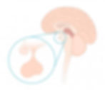
What is the Pituitary Gland?Located at the base of the brain, the pituitary gland controls hormones affecting growth, metabolism, and stress response. Just above it lies the optic chiasm, where nerve fibers from each eye cross and transmit visual information to the brain.
Suggested Image: Diagram showing pituitary gland and optic chiasmCaption: The pituitary gland lies directly beneath the optic chiasm, making vision loss a possible symptom of pituitary lesions.
What Are Pituitary Lesions?Pituitary lesions are abnormal growths (often benign tumors) that develop near or on the gland. When they expand, they can compress the optic chiasm, leading to chiasmal injury and pituitary lesion vision loss.
Common Symptoms of Pituitary Lesion Vision Loss
Patients may experience subtle or sudden visual changes, including:
Blurred or dim vision
Loss of peripheral (side) vision, often in both eyes (bitemporal hemianopsia)
Headaches or hormonal imbalance symptoms (fatigue, weight changes, menstrual irregularities)
In many cases, visual symptoms are the first sign of a pituitary tumor—making prompt eye exams critical.
Causes and Risk Factors
Most pituitary lesions are adenomas—noncancerous tumors. While the exact cause remains unclear, contributing factors may include:
Genetic predisposition (e.g., MEN1 syndrome)
Hormonal imbalances
Rarely, metastases or inflammatory diseases
How Pituitary Lesion Vision Loss is Diagnosed
Dr. Narain uses a combination of cutting-edge retinal imaging and visual field testing to detect signs of chiasmal compression. Key diagnostic tools include:
OCT (Optical Coherence Tomography): Detects thinning in the retinal nerve fiber layer and ganglion cell layer—early signs of chiasmal injury.
Visual Field Testing: Identifies characteristic patterns of vision loss.
Neuroimaging (MRI): Ordered when eye findings suggest a central cause.
Visual Evoked Potentials (VEP): Enables objective assessment of optic nerve function and can reveal abnormalities linked to tumor compression—technology few clinics offer.
Treatment Options for Pituitary Lesions
Treating pituitary lesions often requires a multidisciplinary approach involving endocrinologists and neurosurgeons. Treatment may include:
Observation (for small, stable lesions)
Surgical removal via a transsphenoidal approach
Medical therapy for hormone-secreting tumors
Radiation therapy if surgery isn’t an option
At South Bay Retina, we monitor visual recovery or progression after neurosurgery using OCT and field tests—ensuring patients receive coordinated, personalized care.
Advanced Neuro-Ophthalmic Care at South Bay Retina
Dr. Narain’s protocol emphasizes early detection, even before MRI changes appear. Our clinic offers:
Detailed visual field and OCT evaluations
Ganglion Cell Layer analysis to detect damage sooner than conventional methods
Ongoing collaboration with neurologists and neurosurgeons for seamless patient care
Takeaway Message: Your Eyes May Reveal a Hidden Problem
Pituitary lesions don’t always announce themselves loudly—but your eyes might. If you're noticing peripheral vision loss, persistent headaches, or unexplained hormonal symptoms, consult with a retina specialist immediately.
At South Bay Retina, we’re not just treating eyes—we’re protecting lives through early detection and expert care.
Watch the Video here:
Listen to the Podcast here: