.png)
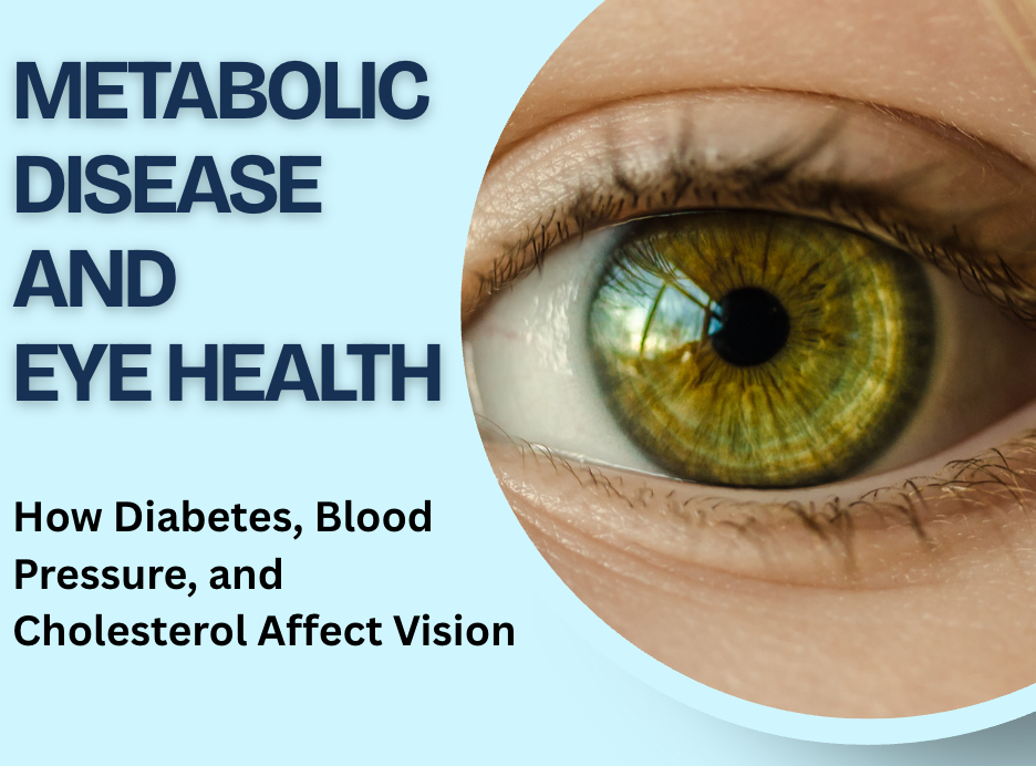
Metabolic Disease and Eye Health: How Diabetes, Blood Pressure, and Cholesterol Affect Vision

Blood sugar, blood pressure, and cholesterol are usually discussed in relation to the heart, kidneys, or risk of stroke.
They also matter to your eyes.
The retina contains an intricate network of tiny blood vessels responsible for supplying oxygen and nutrients to the cells involved in vision. Conditions such as diabetes, hypertension, and high cholesterol place stress on this circulation.
When several of these conditions occur together, doctors often refer to a broader pattern of metabolic dysfunction or, in certain circumstances, metabolic syndrome.
The eyes do not exist separately from the rest of the body. Changes affecting blood vessels throughout the body often appear in the retina as well.
Understanding this connection helps explain why protecting your metabolic health also supports long-term vision.
1. What does metabolic health mean?
Metabolic health describes how effectively the body regulates processes including:
blood sugar
blood pressure
cholesterol and triglycerides
insulin response
body fat distribution
Problems in these areas frequently overlap.
For example, someone with insulin resistance might also have high blood pressure and abnormal cholesterol levels.
A specific combination of metabolic risk factors is known as metabolic syndrome.
These conditions matter because they influence blood-vessel health throughout the body, including the tiny vessels inside the retina.
2. Why is the retina especially sensitive to blood-vessel health?
The retina is highly active tissue.
Its cells constantly process light and send visual information toward the brain through the optic nerve.
This activity requires a reliable supply of oxygen.
The retina therefore contains an extensive network of arteries, veins, and microscopic capillaries.
Damage to these vessels leads to:
leakage
bleeding
swelling
reduced blood flow
loss of oxygen
abnormal new blood-vessel growth
Different metabolic conditions affect these vessels in different ways.
When several risk factors occur together, their effects often overlap.
3. Diabetes damages tiny retinal blood vessels
Diabetes is one of the clearest examples of the connection between metabolic health and vision.
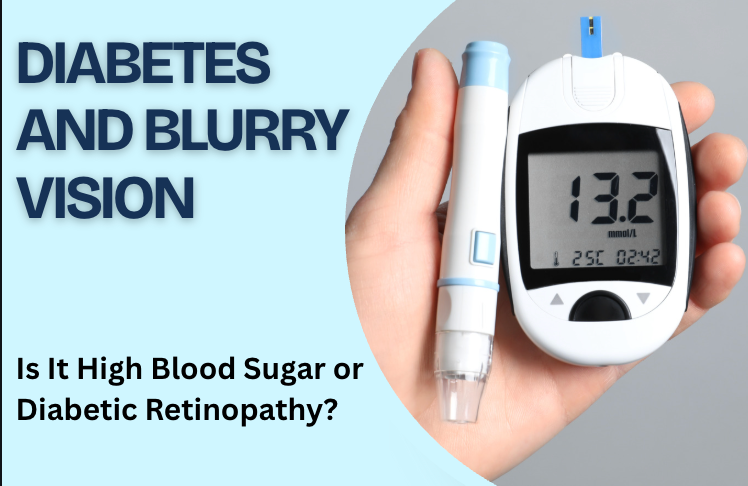
Long-term elevated blood sugar damages retinal capillaries.
The vessels gradually become more likely to:
leak
bleed
become blocked
form small bulges
lose normal circulation
These changes are known as diabetic retinopathy.
Early diabetic retinopathy often develops without noticeable symptoms.
As disease progresses, vision problems include blur, floaters, dark areas, or reduced visual detail.
Good vision today does not necessarily mean diabetes has had no effect on the retina.
4. Diabetes also leads to macular swelling
The macula is the central part of the retina responsible for sharp detailed vision.
When damaged diabetic blood vessels leak fluid into this area, the macula becomes swollen.
This is called diabetic macular edema, or DME.
Symptoms often include:
blurry central vision
wavy lines
difficulty reading
reduced fine detail
colors appearing less vivid
OCT imaging shows this retinal fluid in detailed cross-section.
For patients with vision-affecting DME, retinal injections are one of the main modern treatments.
5. High blood pressure affects retinal arteries
High blood pressure places additional stress on blood-vessel walls.
Over time, retinal arteries become narrower and less flexible.
Doctors refer to characteristic retinal changes associated with hypertension as hypertensive retinopathy.
An examination sometimes shows:
narrowed retinal arteries
changes where arteries cross veins
small retinal hemorrhages
cotton-wool spots
retinal leakage
Mild hypertensive retinopathy often produces no symptoms.
Severe hypertension leads to much more significant retinal injury and sometimes affects the optic nerve as well.
6. High blood pressure also raises the risk of retinal vessel blockages
Hypertension affects more than the appearance of retinal arteries.
It is also an important risk factor for retinal vascular occlusions.
A retinal vein occlusion occurs when blood has difficulty draining away from the retina through a vein.
This often leads to:
retinal bleeding
macular swelling
blurry vision
distorted vision
A retinal artery occlusion occurs when an artery supplying the retina becomes blocked.
This often causes sudden painless loss of vision and requires emergency medical assessment.
Healthy blood pressure is therefore important for both chronic retinal health and the risk of acute vascular events.
7. High cholesterol affects the arteries supplying your eyes
High LDL cholesterol contributes to atherosclerosis.
Atherosclerosis means fatty plaque gradually develops within artery walls.
This narrows blood vessels and increases the risk of material breaking away and traveling through the bloodstream.
A tiny cholesterol-containing embolus sometimes becomes lodged inside a retinal artery.
In some patients, an eye doctor sees this during a dilated retinal examination before the patient notices any vision problem.
A larger blockage interrupts retinal blood flow and results in retinal artery occlusion.
8. These risk factors often occur together
Diabetes, hypertension, and abnormal cholesterol frequently overlap.
This matters because their effects on blood vessels are not completely independent.
Someone with diabetes and well-controlled blood pressure faces a different risk profile from someone with diabetes, persistent hypertension, high cholesterol, and smoking exposure.
The National Eye Institute specifically notes that high blood pressure or high cholesterol alongside diabetes increases the risk of diabetic retinopathy.
This is one reason retinal health needs to be viewed within the bigger picture of metabolic and cardiovascular health.
9. Metabolic health also affects the optic nerve
The retina is not the only structure influenced by systemic disease.
The optic nerve carries visual information from the retina toward the brain.
Diabetes is associated with a higher risk of several eye conditions beyond diabetic retinopathy, including glaucoma. High blood pressure also affects circulation supplying the optic nerve and other ocular tissues.
A comprehensive eye examination therefore looks beyond the retina alone.
Eye pressure, the optic nerve, lens, macula, and peripheral retina all provide useful information.
10. Vision changes are not always the first warning
One of the most important things about metabolic eye disease is how quietly it often starts.
Early diabetic retinopathy often has no symptoms.
Mild hypertensive retinal changes also often go unnoticed.
High cholesterol usually produces no eye symptoms unless vascular disease causes a more significant retinal event.
This means waiting for vision to become blurry is not an effective way to monitor eye health.
For people with diabetes or high blood pressure, dilated examinations are especially important because retinal changes are often visible before the patient notices them. NEI notes that most people with diabetes or high blood pressure need a dilated examination at least annually, with individual schedules set by their doctors.
11. Sudden blurry vision is different from gradual change
Metabolic conditions produce both chronic and acute eye problems.
A gradual change in vision sometimes results from:
diabetic macular edema
cataracts
changing prescription
chronic retinal disease
Sudden vision loss raises different concerns.
Possible causes include:
retinal artery occlusion
retinal vein occlusion
vitreous hemorrhage
retinal detachment
other vascular or neurologic events
A sudden dramatic change in one eye should not be dismissed as “blood sugar” or “blood pressure.”
Prompt assessment is important.
12. Why controlling blood sugar matters
For someone with diabetes, glucose control remains one of the most important parts of protecting retinal health.
Persistent high blood sugar drives damage to retinal capillaries and contributes to inflammation, leakage, poor circulation, and abnormal blood-vessel growth.
Keeping glucose within an individualized target reduces the risk of diabetic retinal disease progressing.
This works alongside regular retinal monitoring rather than replacing it.
Someone with excellent blood sugar control still needs the eye examinations recommended for their individual situation.
13. Blood pressure control matters just as much
Blood pressure affects the structural health of small vessels.
When pressure remains elevated for years, arteries become stiffer and more damaged.
For people with diabetes, hypertension adds another source of stress to retinal circulation.
Managing blood pressure therefore supports both systemic health and the retina.
This involves following the treatment plan provided by the clinician managing your hypertension, including prescribed medication and lifestyle recommendations.
14. Cholesterol is another piece of the picture
Cholesterol often receives less attention in conversations about eyesight than blood sugar.
It still matters.
High cholesterol contributes to atherosclerosis and vascular disease.
For someone with diabetes, cholesterol control also forms part of reducing the risk of diabetic retinal complications. NEI specifically recommends managing diabetes, blood pressure, and cholesterol as part of protecting vision in diabetic retinopathy.
A retina specialist does not replace your primary-care physician or cardiologist, but retinal findings sometimes provide another reason to review cardiovascular risk factors.
15. Smoking adds another vascular stress
Smoking damages blood vessels and contributes to cardiovascular disease.
It also raises the risk of several important eye conditions.
For someone who already has diabetes, hypertension, or high cholesterol, smoking adds another modifiable risk factor affecting long-term health.
Avoiding tobacco supports the same circulation relied upon by the retina, optic nerve, heart, and brain.
NEI includes stopping smoking and staying physically active among its recommendations for maintaining healthy vision and reducing conditions such as diabetes, high blood pressure, and high cholesterol.
16. Exercise supports eye health indirectly
Exercise does not treat a retinal tear or remove macular edema.
Its benefit comes through broader health.
Regular physical activity supports:
blood sugar control
blood pressure management
cardiovascular fitness
healthy cholesterol levels
weight management
These factors reduce the burden of conditions responsible for many retinal vascular problems.
Protecting your vision therefore extends beyond what happens during an eye appointment.
17. What does a retina specialist look for?
During a dilated retinal examination, a retina specialist looks for signs including:
retinal hemorrhages
leaking vessels
blocked vessels
abnormal vessel growth
macular swelling
areas of poor retinal circulation
cholesterol emboli
changes associated with hypertension
Several imaging tests provide additional detail when needed.
18. OCT detects retinal swelling
Optical coherence tomography, or OCT, creates detailed cross-sectional images of the retina.
It is particularly useful for detecting:
diabetic macular edema
swelling after retinal vein occlusion
fluid related to macular disease
structural changes in the macula
OCT also allows doctors to compare retinal thickness and fluid between appointments.
This helps guide decisions about treatment.
19. Retinal photography provides a record of blood-vessel health
Retinal photographs document the appearance of the retina and its circulation.
They show:
hemorrhages
abnormal vessels
vascular narrowing
retinal changes associated with diabetes
changes over time
For metabolic and vascular disease, being able to compare current findings with earlier photographs is especially useful.
20. Your eye examination sometimes reveals a wider health problem
The retina is one of the few places where small blood vessels are directly visible.
As a result, eye doctors sometimes identify vascular abnormalities in patients who feel otherwise well.
Finding retinal changes does not diagnose every cardiovascular or metabolic condition.
It does sometimes prompt further evaluation of:
blood pressure
blood sugar
cholesterol
cardiovascular risk
The eye examination and medical assessment complement each other.
When should you schedule a retinal examination?
Arrange an eye examination if you have:
diabetes
high blood pressure
several metabolic risk factors
persistent blurry vision
distorted or wavy vision
unexplained changes between the two eyes
new floaters
a history of retinal vascular disease
If you already have diabetes or hypertension, follow the examination schedule recommended by your medical and eye-care teams.
Seek urgent care for sudden symptoms
Seek prompt medical attention for:
sudden loss of vision in one eye
a new curtain or shadow
a sudden large increase in floaters
sudden dense visual haze
abrupt loss of peripheral vision
sudden vision loss with weakness, facial drooping, or difficulty speaking
These symptoms sometimes indicate an acute retinal or neurologic vascular event.
What this means for your eyes
Blood sugar, blood pressure, and cholesterol are not separate from eye health.
They influence the small blood vessels responsible for keeping retinal tissue healthy.
Diabetes damages retinal capillaries and leads to diabetic retinopathy and macular edema. Hypertension changes retinal blood vessels and increases the risk of vascular occlusion. High cholesterol contributes to atherosclerosis and retinal artery blockages.
When these conditions occur together, managing the whole metabolic picture becomes especially important.
Regular eye examinations, alongside good control of blood sugar, blood pressure, cholesterol, and other cardiovascular risk factors, provide a stronger foundation for protecting vision over the long term.
Book with South Bay Retina
If you have diabetes, high blood pressure, high cholesterol, or unexplained changes in your vision, schedule a retinal evaluation with South Bay Retina.
A comprehensive dilated retinal examination and appropriate imaging help identify vascular changes, macular swelling, and other retinal problems before they have a larger impact on your sight.

Comments