.png)
Diabetic Macular Edema vs. Diabetic Retinopathy: What’s the Difference?

Diabetic retinopathy and diabetic macular edema are closely connected, but they do not mean exactly the same thing.
Diabetic retinopathy is a broad condition caused by diabetes damaging blood vessels throughout the retina.
Diabetic macular edema, often shortened to DME, develops when damaged retinal blood vessels leak fluid into the macula, causing the center of the retina to swell.
You can have diabetic retinopathy without diabetic macular edema. DME develops as a complication of diabetic retinopathy and may occur at different stages of the disease.
Understanding the difference matters because each affects vision differently and treatment decisions depend on what is happening inside the retina.

1. What is diabetic retinopathy?
The retina is the light-sensitive tissue lining the back of the eye.
It contains a network of tiny blood vessels responsible for delivering oxygen and nutrients to retinal cells.
Over time, diabetes damages these vessels.
They may:
weaken
leak fluid
bleed
close off
develop small bulges
trigger abnormal new blood-vessel growth
These changes are collectively known as diabetic retinopathy.
The disease often begins without noticeable symptoms. Someone might see clearly while retinal blood-vessel damage is already developing.
This is why regular diabetic eye examinations matter even when vision seems normal.
2. What is diabetic macular edema?
The macula is the small central part of the retina responsible for sharp, detailed vision.
You use it when:
reading
driving
recognizing faces
using a phone
seeing fine detail
When diabetes damages retinal blood vessels, they sometimes begin leaking fluid.
If this fluid collects in the macula, the retinal tissue becomes swollen.
This is called diabetic macular edema.
DME often produces central blur or distortion because the macula is directly responsible for detailed central vision.
3. The easiest way to understand the difference
Think of diabetic retinopathy as the underlying retinal blood-vessel disease.
Diabetic macular edema is one specific complication of that disease.
In simple terms:
Diabetic retinopathy = diabetes has damaged retinal blood vessels.
Diabetic macular edema = damaged vessels are leaking enough fluid to swell the macula.
Not everyone with diabetic retinopathy develops DME.
However, diabetic macular edema occurs because diabetic retinal blood vessels have become damaged and leaky.
4. Diabetic retinopathy has different stages
Diabetic retinopathy generally progresses through two broad stages.
Nonproliferative diabetic retinopathy
This is the earlier stage.
Doctors may see:
tiny blood-vessel bulges called microaneurysms
small retinal hemorrhages
leaking fluid
deposits within the retina
areas of reduced blood flow
The severity ranges from mild to severe.
Many people have no symptoms during this stage.
Proliferative diabetic retinopathy
This is the more advanced stage.
When areas of the retina receive too little oxygen, the eye releases signals encouraging new blood vessels to grow.
Unfortunately, these vessels are fragile and abnormal.
They sometimes:
bleed into the vitreous
produce sudden dark floaters
cause vitreous hemorrhage
form scar tissue
pull on the retina
lead to tractional retinal detachment
Proliferative diabetic retinopathy threatens sight even when macular edema is not present.
5. DME can occur at different stages
A common misconception is that diabetic macular edema only appears at the final stage of diabetic retinopathy.
That is not true.
DME develops when damaged retinal vessels leak into the macula, and this can happen during different stages of diabetic retinopathy.
Someone might have nonproliferative diabetic retinopathy and significant macular edema.
Another person might have advanced proliferative disease without major macular swelling.
This is why doctors assess both the stage of diabetic retinopathy and the condition of the macula.
They answer two different questions.
6. The symptoms are different
Diabetic retinopathy often produces no symptoms early on.
As disease progresses, symptoms might include:
blurry vision
floaters
dark spots
areas of missing vision
poor night vision
sudden vision changes from retinal bleeding
DME more specifically affects central vision.
Typical symptoms include:
blurry central vision
straight lines looking wavy
difficulty reading
reduced detail
colors looking washed out
objects appearing different sizes between the two eyes
A patient sometimes has both sets of symptoms if diabetic retinopathy and macular edema are present together.
7. Why diabetes damages retinal blood vessels
High blood sugar over time affects blood vessels throughout the body.
The tiny retinal vessels are particularly vulnerable.
Their walls may weaken, and the normal barrier controlling movement of fluid between the bloodstream and retinal tissue begins to break down.
This leads to leakage.
Other retinal vessels may gradually close.
The combination of leakage and reduced blood flow drives much of the damage seen in diabetic retinopathy.
The longer someone has diabetes, the greater their risk of diabetic eye disease.
Blood pressure and cholesterol also influence retinal vascular health.
8. Why DME makes vision blurry
The macula has a highly organized structure.
Its retinal cells need to remain precisely arranged for detailed vision to remain sharp.
When fluid collects inside the macula, retinal layers become thickened and separated.
The image reaching the brain then loses clarity.
That is why stronger glasses often fail to correct blur caused by diabetic macular edema.
The problem is not simply focusing light differently.
The retina itself is swollen.
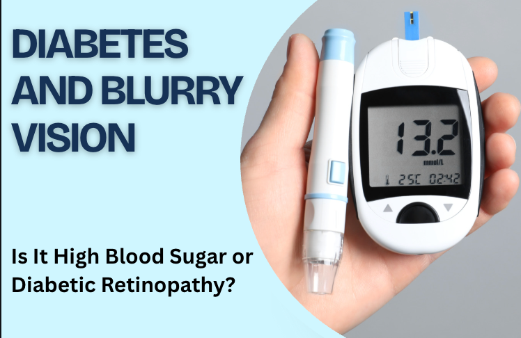
9. Can high blood sugar cause blurry vision without retinopathy?
Yes.
Changes in blood sugar sometimes temporarily affect the lens inside the eye and alter how it focuses.
This creates blurry vision even without new retinal damage.
Vision related to fluctuating blood sugar may change as glucose levels change.
Diabetic macular edema is different.
DME involves physical fluid accumulation inside retinal tissue.
Persistent blur, distortion, or a noticeable difference between the two eyes should therefore not automatically be blamed on blood sugar.
An examination helps determine whether the problem comes from temporary focusing changes or retinal disease.
10. How is diabetic retinopathy diagnosed?
A comprehensive dilated eye examination allows the doctor to look for diabetic retinal changes.
The examination may reveal:
microaneurysms
retinal hemorrhages
leaking vessels
abnormal new vessels
scar tissue
retinal swelling
Retinal photographs are also useful for documenting disease severity and comparing changes over time.
Additional imaging is selected according to what the examination shows.
11. OCT is especially important for DME
Optical coherence tomography, or OCT, produces detailed cross-sectional images of the retina.
For diabetic macular edema, OCT is particularly useful because it shows fluid within retinal layers.
A retina specialist sees:
whether swelling is present
where fluid is located
how thick the macula has become
whether the center of the macula is involved
whether treatment is reducing the fluid
Repeated OCT scans provide an objective way to monitor progress.
A patient might notice improvement in vision while OCT simultaneously shows reduced swelling.
12. What does fluorescein angiography show?
In selected patients, fluorescein angiography provides additional information about retinal circulation.
A fluorescent dye is injected into a vein in the arm and travels through retinal blood vessels.
A special camera then photographs the retina.
This helps the doctor identify:
leaking blood vessels
blocked vessels
areas receiving inadequate blood flow
abnormal new blood-vessel growth
OCT focuses strongly on retinal structure and swelling, while angiography provides more information about blood flow and leakage.
13. How is diabetic macular edema treated?
Treatment depends on the severity of swelling, its effect on vision, and other retinal findings.
Anti-VEGF injections
Anti-VEGF medicines are a major treatment for vision-affecting DME.
Diabetic retinal disease increases levels of VEGF, a protein involved in abnormal vessel permeability and growth.
Anti-VEGF medication reduces this activity.
This helps:
reduce leakage
decrease macular swelling
stabilize vision
improve vision in many patients
Treatment usually involves repeated injections rather than a single procedure.
The schedule changes according to how the retina responds.
14. Are steroids used for DME?
Steroid treatment is useful for selected patients.
Steroids reduce inflammation and retinal swelling.
They may be delivered as injections or longer-lasting implants.
However, steroids also increase the likelihood of:
elevated eye pressure
glaucoma
cataract progression
The retina specialist weighs these risks against the potential benefit.
Anti-VEGF treatment is often used first for center-involving DME affecting vision, with other options selected based on individual circumstances.
15. Is laser still used?
Yes, although its role has changed.
Before modern retinal injections became widely available, laser played a much larger role in DME treatment.
Laser remains useful in selected cases.
Treatment might target areas of retinal leakage outside the very center of the macula.
For proliferative diabetic retinopathy, a different approach called panretinal photocoagulation is often used to treat areas of retina producing signals for abnormal blood-vessel growth.
This highlights another difference between DME and broader diabetic retinopathy.
The treatment chosen depends on which complication is present.
16. How is proliferative diabetic retinopathy treated?
Proliferative diabetic retinopathy focuses on controlling abnormal new blood vessels and reducing the risk of severe bleeding or retinal detachment.
Treatment might include:
anti-VEGF injections
retinal laser treatment
vitrectomy surgery in advanced cases
A patient with both proliferative retinopathy and DME sometimes needs treatment addressing both problems.
The management plan is tailored to the retina rather than treating diabetes-related eye disease as one identical condition.
17. When is vitrectomy needed?
Vitrectomy is retinal surgery used for advanced complications rather than routine mild diabetic retinopathy.
It is considered when problems include:
persistent vitreous hemorrhage
significant scar tissue
traction on the retina
tractional retinal detachment
certain cases involving macular traction
During surgery, the vitreous gel and blood or scar tissue interfering with the retina are removed.
The goal depends on the complication being treated.
18. Does controlling diabetes still matter after retinopathy develops?
Yes.
Eye treatment does not replace diabetes management.
Controlling:
blood sugar
blood pressure
cholesterol
helps reduce the risk of diabetic eye disease progressing.
Diabetes affects retinal blood vessels continuously, so systemic health remains important even when retinal injections or laser treatment are working well.
Patients should continue working with the medical team managing their diabetes alongside their retina specialist.
19. Do people with diabetes need eye exams if they see perfectly?
Yes.
One of the most important facts about diabetic retinopathy is that early disease often has no symptoms.
Clear vision does not prove the retina is unaffected.
A dilated examination identifies changes before the patient notices them.
The National Eye Institute advises people with diabetes to have a comprehensive dilated eye examination at least once a year, with examination frequency adjusted based on individual findings.
People already diagnosed with diabetic retinopathy often need more frequent monitoring.
20. What happens if DME is untreated?
Persistent macular swelling damages the retinal cells responsible for detailed central vision.
Over time, untreated DME results in increasing difficulty with:
reading
driving
recognizing faces
seeing fine detail
Long-standing retinal changes also limit how much vision improves after treatment begins.
Earlier diagnosis provides a better opportunity to control swelling before substantial permanent retinal damage develops.
21. What happens if diabetic retinopathy is untreated?
The risks depend on the stage.
Early changes sometimes remain stable for long periods with good diabetes management and monitoring.
Advanced disease may lead to:
vitreous hemorrhage
severe retinal ischemia
abnormal blood-vessel growth
neovascular glaucoma
scar tissue
tractional retinal detachment
permanent vision loss
This is why retinal monitoring continues even when DME is absent.
Diabetic retinopathy vs. DME at a glance
Diabetic retinopathy
What it is:Diabetes-related damage to blood vessels throughout the retina.
Early symptoms:Often none.
Later problems:Bleeding, abnormal vessels, floaters, retinal scarring, retinal detachment, and vision loss.
Diabetic macular edema
What it is:Fluid leaking into and swelling the macula as a result of diabetic retinal blood-vessel damage.
Common symptoms:Central blur, distortion, wavy lines, reduced detail, and faded colors.
Main concern: Damage to detailed central vision.
A person may have diabetic retinopathy without DME, while DME develops as a complication of diabetic retinal vascular damage.
When should someone with diabetes see a retina specialist?
Schedule an eye examination if you have diabetes and notice:
persistent blurry vision
wavy or distorted central vision
new floaters
dark spots
a noticeable difference between the eyes
difficulty reading
sudden visual haze
reduced central detail
You should also keep recommended diabetic eye examinations even without symptoms.
Retinal disease often begins quietly.
Seek prompt attention for sudden symptoms
Seek urgent eye care for:
a sudden shower of dark floaters
abrupt severe vision loss
a curtain or shadow
sudden loss of peripheral vision
flashes of light with new floaters
sudden dense haze in one eye
Advanced diabetic retinopathy sometimes causes vitreous bleeding or retinal detachment, both of which need timely evaluation.
What this means for your eyes
Diabetic retinopathy and diabetic macular edema are connected, but they describe different parts of diabetic eye disease.
Diabetic retinopathy refers broadly to damage affecting retinal blood vessels.
Diabetic macular edema occurs when damaged vessels leak fluid into the macula and cause central retinal swelling.
You can have diabetic retinopathy without DME. DME is one important complication of diabetic retinopathy.
Regular dilated examinations help identify diabetic retinal changes before symptoms appear. OCT imaging then provides detailed information when macular swelling is suspected.
Modern treatments such as anti-VEGF injections have significantly changed how diabetic macular edema and diabetic retinopathy are managed, but early detection remains one of the most important ways to protect vision.
Book with South Bay Retina
If you have diabetes, persistent blurry vision, or have been told you have diabetic retinopathy or diabetic macular edema, schedule a retinal evaluation with South Bay Retina.
A comprehensive dilated retinal examination and OCT imaging help determine the stage of diabetic retinal disease, identify macular swelling, and guide an appropriate monitoring or treatment plan.


Comments