.png)
Macular Hole: Symptoms, Causes, Surgery, and Recovery

A small opening in the center of the retina can have a surprisingly large effect on vision.
A macular hole forms in the macula, the part of the retina responsible for sharp central vision. This is the vision you rely on for reading, recognizing faces, driving, and seeing fine detail.
Early symptoms often include mild blur or straight lines beginning to look wavy. As the hole progresses, a person might notice a blurry, dark, or missing area directly in the center of vision.
Macular holes are most common in adults over 60 and often develop because of age-related changes in the vitreous gel inside the eye.
Small holes sometimes close or remain stable without surgery. Larger or symptomatic full-thickness macular holes often require a retinal procedure called a vitrectomy.
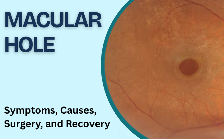
1. A macular hole forms in the center of the retina
The retina is the light-sensitive tissue lining the back of the eye.
At its center is the macula.
Although the macula occupies only a small area, it provides the detailed central vision needed for tasks such as:
reading
recognizing faces
driving
using a phone
seeing fine details
distinguishing colors
At the very center of the macula lies the fovea, where vision is sharpest.
A macular hole develops when the retinal tissue in this central area separates enough to create an opening.
Because peripheral retina remains intact, a macular hole does not usually eliminate all vision. Instead, it primarily affects what you see directly in front of you.
2. Early symptoms often begin with blur or distortion
Macular holes often develop gradually.
At first, the symptoms are easy to dismiss.
You might notice:
central vision becoming slightly blurry
straight lines looking bent or wavy
difficulty reading small print
letters appearing distorted
faces looking less clear
one eye seeing differently from the other
Some people notice the problem while looking at a door frame, window frame, spreadsheet, or grid.
The medical term for visual distortion is metamorphopsia.
As the hole becomes larger, the change often becomes more noticeable.
3. A dark or missing central spot sometimes develops
More advanced macular holes often produce a small area of missing or severely blurred central vision.
For example, you might look directly at someone's face and see the outline of the head but struggle to see the eyes or nose clearly.
While reading, parts of words might disappear.
This central blind or blurry area is known as a central scotoma.
Peripheral vision generally remains available, so someone with a macular hole often still sees objects beside them.
That pattern is different from diseases which primarily affect peripheral vision.
4. Age-related vitreous changes are the most common cause
Most macular holes develop as part of changes inside the eye associated with ageing.
The center of the eye contains a clear gel called the vitreous.
Earlier in life, the vitreous has a firmer gel-like structure. Over time, it becomes more liquid and gradually separates from the retina.
Usually, this separation happens without damaging the macula.
In some eyes, however, part of the vitreous remains firmly attached to the center of the retina.
As the vitreous pulls away, it creates traction on the macula.
If the pulling becomes strong enough, the central retinal tissue stretches and an opening forms.
5. Vitreomacular traction often comes before a macular hole
Before a full macular hole develops, OCT imaging sometimes shows the vitreous pulling on the macula.
This is called vitreomacular traction.
The traction alters the normal shape of the central retina.
Some cases resolve as the vitreous finishes separating from the retina.
In others, continued traction progresses into a full-thickness macular hole.
This is one reason OCT imaging is so useful. It shows not only whether a hole exists, but also the relationship between the vitreous and the macula.
6. Age is an important risk factor
Macular holes occur most often in people over 60.
The risk rises with age because vitreous separation becomes increasingly common.
Other factors associated with macular holes include:
high myopia, or strong nearsightedness
previous eye trauma
previous eye surgery
certain retinal conditions
Most macular holes still develop without an injury or another obvious retinal disease.
They are often described as idiopathic macular holes, meaning they arise without a separate identifiable cause beyond age-related vitreous changes.
7. High myopia changes the structure of the eye
People with high myopia have eyes that are longer than average.
This stretches the retina and other tissues at the back of the eye.
Those structural differences increase the risk of several retinal problems, including macular holes.
A macular hole in a highly myopic eye sometimes behaves differently from a typical age-related hole, especially if additional retinal thinning or detachment is present.
People with strong nearsightedness should report new central distortion or missing vision promptly.
8. Eye trauma sometimes causes a macular hole
A strong blow to the eye sometimes creates a traumatic macular hole.
This is more common in younger people than ordinary age-related macular holes.
Possible examples include injuries from:
sports
falls
workplace accidents
being struck by an object
Some traumatic macular holes close without surgery, while others persist and require treatment.
A retina specialist evaluates the size of the hole, retinal anatomy, symptoms, and other damage before deciding on management.
9. Macular hole and macular pucker are different
The terms sound similar, and the symptoms often overlap.
A macular pucker develops when a thin membrane forms over the retina and wrinkles the macular surface.
A macular hole is an opening in the central retinal tissue itself.
Both often cause:
wavy lines
blurry central vision
distorted reading
difficulty with fine detail
The difference becomes clear on OCT imaging.
This distinction matters because the retinal anatomy and surgical approach differ.
10. Macular hole is not macular degeneration
Macular holes are also different from age-related macular degeneration, or AMD.
Both affect central vision, and both occur more frequently in older adults.
However, they are separate conditions.
AMD involves degeneration of retinal tissues and, in wet AMD, abnormal leaking blood vessels.
A macular hole results from a structural opening in the center of the macula.
Similar symptoms do not mean the diseases are the same.
A retinal examination and OCT usually distinguish them quickly.
11. OCT is the key test for diagnosing a macular hole
Optical coherence tomography, or OCT, provides detailed cross-sectional images of the retina.
The scan shows the retina in layers and clearly demonstrates whether an opening exists in the macula.
OCT helps the retina specialist assess:
the size of the hole
whether it extends through the full retinal thickness
vitreous traction
surrounding retinal swelling
the shape of the hole
changes during follow-up
whether the hole has closed after surgery
The test is non-invasive and usually takes only a short time.
A comprehensive dilated retinal examination is also performed to assess the rest of the retina.
12. Some small macular holes are monitored
Not every macular hole goes directly to surgery.
Very small or early holes with limited symptoms sometimes receive close observation.
The decision depends on factors including:
hole size
OCT appearance
degree of vitreous traction
visual symptoms
whether the hole is progressing
Regular OCT scans show whether the macular anatomy is improving, remaining stable, or worsening.
For a larger full-thickness hole affecting vision, surgery is usually the standard treatment.
13. Vitrectomy is the main surgery for a macular hole
The standard operation is called a pars plana vitrectomy.
During surgery, the retina surgeon removes the vitreous gel from inside the eye.
This eliminates the traction pulling on the macula.
The surgeon also often removes a very thin layer from the retinal surface called the internal limiting membrane.
Removing this tissue reduces residual traction around the hole and supports closure.
A gas bubble is then placed inside the eye.
The gas acts as temporary internal support while the edges of the macular hole come together and heal.
14. The gas bubble gradually disappears
The gas bubble does not stay permanently.
Over time, the eye naturally absorbs the gas and replaces it with its own fluid.
While the bubble remains inside the eye, vision is significantly blurred.
Patients often describe seeing a moving line representing the top of the bubble.
As the bubble shrinks, this line moves lower in the visual field until the gas disappears completely.
Your surgeon will tell you how long the specific gas used is expected to remain.
15. Positioning after surgery depends on the individual case
Some patients are instructed to maintain a particular head position after macular hole surgery, often including periods of face-down positioning.
This helps keep the gas bubble against the macula.
The duration depends on:
the size of the hole
the surgical technique
the type of gas
the surgeon's postoperative plan
Not every patient receives identical positioning instructions.
Follow the instructions from your retinal surgeon rather than using a general positioning schedule found online.
16. Flying is unsafe while a gas bubble remains in the eye
This is one of the most important instructions after surgery involving intraocular gas.
Do not fly while the gas bubble remains inside the eye.
Lower cabin pressure at altitude allows the gas to expand.
Expansion inside the closed eye raises eye pressure rapidly and creates a serious risk to vision.
Travel to significantly higher elevations also requires discussion with your surgeon.
Patients with an intraocular gas bubble must also tell other doctors and anesthesiologists before medical or dental procedures because nitrous oxide anesthesia is unsafe while intraocular gas remains present.
Your retina surgeon will tell you when these restrictions have ended.
17. Vision recovery takes time
Vision does not return immediately after macular hole surgery.
At first, the gas bubble itself makes sight blurry.
After it disappears, retinal recovery continues.
Improvement often develops gradually over weeks or months.
Factors influencing the final visual result include:
size of the macular hole
how long it was present
retinal condition before surgery
whether the hole closes successfully
other eye diseases
Surgery often improves central vision and distortion, but perfect vision is not guaranteed.
Earlier treatment generally offers a better opportunity for visual recovery than waiting until a large hole has been present for a long time.
18. Macular hole surgery has a high closure rate
Modern vitrectomy successfully closes most macular holes.
The American Society of Retina Specialists reports anatomical closure rates above 90% with vitrectomy.
Successful closure does not always mean vision returns completely to normal.
Some retinal cells have already been disrupted before surgery, especially when the hole has been present for a longer period.
The primary goals are to close the hole, improve visual function where possible, and prevent continued central vision loss.
19. Cataracts often progress after vitrectomy
One important consideration for patients who still have their natural lens is cataract progression.
Vitrectomy often speeds up cataract formation, particularly in older adults.
A cataract clouds the natural lens and produces symptoms such as:
blurry vision
glare
difficulty driving at night
faded colors
Some patients therefore need cataract surgery after recovering from vitrectomy.
Your retina surgeon and cataract surgeon coordinate the timing based on your individual eyes.
20. Other surgical risks are uncommon but important
As with any eye operation, vitrectomy has potential complications.
These include:
retinal tear
retinal detachment
infection
bleeding
increased eye pressure
cataract progression
failure of the hole to close
reopening of a previously closed hole
Your retina surgeon discusses these risks before surgery and compares them with the expected benefit of treatment.
21. A macular hole in one eye increases attention to the other eye
Most patients initially develop a macular hole in one eye.
The National Eye Institute reports that roughly 1 in 10 people who develop a macular hole in one eye later develop one in the other eye.
This does not mean the second eye will inevitably be affected.
It does mean new distortion or central blur in the other eye deserves evaluation.
Regular monitoring is particularly useful when OCT shows persistent vitreous attachment in the fellow eye.
When to see a retina specialist
Arrange a retinal evaluation if you notice:
straight lines becoming wavy
new central blur in one eye
letters appearing broken or missing
a small central blind spot
faces looking distorted
increasing difficulty reading with one eye
a noticeable difference between your two eyes
These symptoms overlap with several other macular conditions, so retinal imaging is important for diagnosis.
Symptoms requiring faster attention
A macular hole usually develops gradually.
Seek prompt eye care if you experience:
sudden major vision loss
new flashes of light
a sudden increase in floaters
a curtain or shadow over vision
abrupt peripheral vision loss
severe eye pain with reduced vision
These symptoms suggest a different retinal or eye emergency.
What this means for your eyes
A macular hole is a small opening in the center of the retina, but its location means it has a large effect on detailed vision.
The first signs often include wavy lines, central blur, and difficulty reading. As the hole progresses, a dark or missing central area sometimes appears.
OCT imaging shows the size and structure of the hole in detail.
Small early holes sometimes receive observation. Symptomatic full-thickness holes are usually treated with vitrectomy, membrane peeling, and a temporary gas bubble.
Recognizing new central distortion early gives your retina specialist the opportunity to diagnose the problem before additional central vision is lost.
Book with South Bay Retina
If straight lines look wavy, the center of your vision appears blurry, or you have been diagnosed with a macular hole, schedule a retinal evaluation with South Bay Retina.
A comprehensive retinal examination and OCT scan help determine the size and stage of the macular hole and whether monitoring or surgery is appropriate.



Comments